
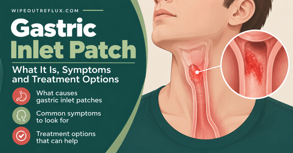
A gastric inlet patch is a small island of stomach lining in the wrong place, at the very top of the esophagus, just below the throat. It’s usually something you’re born with, and it’s more common than most people realize: depending on how carefully the endoscopist looks, it’s found in somewhere between 1 in 100 and 1 in 10 people having an endoscopy.
Here’s why it matters. Stomach lining makes acid, and an inlet patch can do the same, right next to your throat. That means it can, in some people, cause throat symptoms that look exactly like LPR (globus, mucus, throat clearing, a sore or burning throat) without any reflux coming up from the stomach at all.
Whether it actually causes symptoms is where doctors disagree. Many gastroenterologists call an inlet patch “incidental” and move on, and most people who have one never notice it. Others, backed by a small but striking trial, believe it’s an overlooked cause of stubborn throat symptoms. If you’ve been told your scope showed an inlet patch and nobody explained what it means, this article sets out both sides so you can have a better conversation with your doctor.
Key Takeaways
- A gastric inlet patch (also called a cervical inlet patch, esophageal inlet patch, or heterotopic gastric mucosa) is a patch of stomach-type lining in the upper esophagus.
- It’s usually present from birth and is found in roughly 1% to 10% of endoscopies. Many are missed because they sit in an area that’s easy to pass quickly.
- Most inlet patches cause no symptoms, which is why they’re often described as incidental.
- Some patches produce acid and mucus right below the throat, and can cause globus, throat clearing, mucus, hoarseness or a sore throat that mimic LPR.
- In a small sham-controlled trial, burning away inlet patches improved globus in 82% of patients, compared with 0% after a sham procedure.
- An inlet patch is not considered precancerous in the way Barrett’s esophagus is. Cancer arising in one is exceedingly rare.
- Options for a symptomatic patch include acid suppression and, for persistent cases, endoscopic ablation (argon plasma coagulation or radiofrequency ablation).
What Is a Gastric Inlet Patch?
The lining of your esophagus is normally made of flat, tough cells (squamous cells) designed to handle food passing over them. The lining of your stomach is completely different: it’s glandular tissue that produces acid, mucus and digestive enzymes.
An inlet patch is a piece of that stomach-type lining sitting in the upper esophagus, usually just below the upper esophageal sphincter (the muscle ring at the top of your food pipe). On a camera, it looks like a velvety, salmon-pink or red patch with a clear border against the paler esophageal lining. Patches range from a few millimeters across to, rarely, a ring all the way around the esophagus, and some people have two or more.
Crucially, these patches often contain the same working cells as your stomach. In early research, nearly all patches contained both parietal cells, which make acid, and chief cells, which make pepsinogen, the precursor to pepsin [Jabbari et al., Gastroenterology, 1985]. Acid secretion from an inlet patch has been directly recorded on pH monitoring, and it resolved with a PPI [Galan et al., Gastroenterology, 1998].
If you want to understand where the patch sits relative to the throat, my article on the upper esophageal sphincter explains the anatomy.
Inlet patch of esophagus: the different names
You might see any of these on an endoscopy report, and they all mean the same thing:
- Gastric inlet patch
- Cervical inlet patch (CIP)
- Esophageal inlet patch, or inlet patch of the esophagus
- Heterotopic gastric mucosa (HGM), or heterotopic gastric mucosa of the upper esophagus
- Ectopic gastric mucosa
How Common Is It, and Why Is It So Often Missed?
Inlet patches are surprisingly common. In one study of 634 consecutive patients, 10% had one [Borhan-Manesh et al., Gut, 1991]. Other studies report lower figures, and the difference mostly comes down to how carefully the endoscopist looks.
An Italian study showed this neatly. An endoscopist who was specifically looking for inlet patches found them in 2.27% of patients. Another endoscopist, who knew about them but didn’t know he was part of a study, found them in just 0.29% [Maconi et al., European Journal of Gastroenterology & Hepatology, 2000]. That’s nearly an eightfold difference from awareness alone.
The reason is simple. The patch sits just below the upper sphincter, an area the scope passes through quickly on the way in and often on the way out. If nobody slows down and looks, it isn’t seen. So if you’ve had a scope that “found nothing”, it’s possible a small patch was simply not looked for. My guide to endoscopy for acid reflux explains what the procedure covers.
The Big Disagreement: Incidental Finding or Hidden Cause?
This is the heart of the issue, and it explains why one doctor might shrug at your inlet patch while another takes it seriously.
The “incidental” view
Most people with an inlet patch have no symptoms at all. An influential classification published in 2004 puts these asymptomatic patches in the first category (HGM I) and states that they need neither treatment nor surveillance [von Rahden et al., American Journal of Gastroenterology, 2004]. In the Italian prevalence study, inlet patches were only weakly linked to any specific symptom [Maconi et al., European Journal of Gastroenterology & Hepatology, 2000].
From this perspective, a patch is a harmless developmental quirk, and because so many people have one without symptoms, finding a patch in someone with throat symptoms doesn’t prove it’s the cause. That’s a fair point. Your throat symptoms could have another explanation, and the patch could just be along for the ride.
The “hidden cause” view
The same 2004 classification also recognizes a second group (HGM II): people with symptoms such as difficulty or pain swallowing, hoarseness and coughing caused by the patch, without any visible damage [von Rahden et al., American Journal of Gastroenterology, 2004]. Across studies, reported rates of LPR-type symptoms in people with inlet patches range from under 20% to as high as 73%, though most are mild [Chong, World Journal of Gastroenterology, 2013].
The strongest evidence comes from treatment. In a randomized, sham-controlled trial, people with chronic globus and an inlet patch had the patch burned away with argon plasma coagulation or underwent a fake procedure. Symptoms improved in 9 of 11 treated patients (82%) and in none of the 10 sham patients [Bajbouj et al., Gastroenterology, 2009]. That’s a small trial, but a sham control makes the result much harder to dismiss as a placebo effect.
The patch has also made its way into official criteria. Under the Rome IV criteria, an inlet patch should be ruled out before diagnosing functional globus (a lump-in-the-throat feeling with no structural cause) [Rusu et al., Frontline Gastroenterology, 2018].
So who’s right?
Both, really. Most inlet patches are harmless and silent. But a subset clearly do cause symptoms, and the people they affect tend to have had a long, frustrating search for answers. A 2025 study suggests which patches are more likely to cause trouble: larger patches were significantly more common in people with LPR-type symptoms [Wang et al., Gastroenterology Research and Practice, 2025].
The practical question isn’t “do inlet patches cause symptoms?” It’s “is my patch causing my symptoms?” That depends on how big it is, whether your symptoms fit, and whether other causes such as reflux have been properly checked.
How an Inlet Patch Can Cause LPR-Like Symptoms Without Reflux
This is the part I find most interesting, and it’s what I wish someone had explained to the readers who’ve written to me about this.
With LPR, stomach contents travel up the esophagus, past the upper sphincter, and into the throat. With a symptomatic inlet patch, the source is already at the top. It’s a little piece of stomach lining sitting right below your throat, so it doesn’t need anything to travel up. Here’s how it can irritate the area:
- Local acid. Parietal cells in the patch can secrete acid directly into the upper esophagus, right where the esophagus meets the throat.
- Mucus. Stomach lining makes mucus. Inlet patches can secrete both acid and mucus [Dunn et al., Gastrointestinal Endoscopy, 2016], which fits with the thick mucus, phlegm and constant need to clear the throat that many people describe.
- Possibly pepsin. Patches can contain chief cells, which make pepsinogen. Pepsin is the enzyme behind much of the throat damage in LPR, so this is a plausible extra source of irritation, though it hasn’t been studied directly in inlet patches. See what is pepsin.
- Inflammation and H. pylori. Inlet patches can be colonized by H. pylori, especially in people who also have it in their stomach [Gutierrez et al., American Journal of Gastroenterology, 2003], which may add inflammation.
This also explains a frustrating pattern: someone with classic LPR symptoms has a reflux test that comes back fairly normal, or reflux treatment that only partly helps. If the patch is the source, there may be little measurable reflux, and treatments that work on reflux from below (like an alginate raft sitting on top of the stomach) won’t reach acid being made at the top of the esophagus.
Symptoms linked to a symptomatic inlet patch
- Globus, or the feeling of a lump in the throat. See the lump in throat from reflux
- Excess mucus or phlegm in the throat. See phlegm stuck in your throat
- Frequent throat clearing
- Sore or burning throat, often without heartburn. See burning throat with no heartburn
- Hoarseness and chronic cough
- Pain or difficulty swallowing
These overlap almost completely with LPR symptoms, which is exactly why an inlet patch is so easy to overlook.
Is an Inlet Patch Dangerous?
For the vast majority of people, no. That’s worth saying clearly, because “stomach tissue in the esophagus” can sound alarming, especially if you’ve heard of Barrett’s esophagus.
Barrett’s esophagus is a change in the lining of the lower esophagus caused by long-term reflux, and it carries a small increased risk of cancer. An inlet patch is different: it’s usually present from birth, it sits in the upper esophagus, and it isn’t regarded as precancerous in the same way. The 2004 classification describes malignant change as “exceedingly rare” and says that, unlike Barrett’s esophagus, an inlet patch should not be regarded as a precancerous lesion [von Rahden et al., American Journal of Gastroenterology, 2004].
Rare complications do exist. These include ulcers, bleeding, a narrowing (stricture) or web in the upper esophagus, and, very rarely, dysplasia or cancer [Chong, World Journal of Gastroenterology, 2013]. That’s why you should mention new or worsening swallowing difficulty, food sticking, pain on swallowing, weight loss or bleeding to your doctor. If you’re worried about Barrett’s, see is Barrett’s esophagus reversible.
How an Inlet Patch Is Diagnosed
An inlet patch is diagnosed during an upper endoscopy (gastroscopy). The endoscopist sees the characteristic salmon-pink patch just below the upper sphincter and usually takes a small biopsy to confirm it’s stomach-type tissue and check for H. pylori and any abnormal cells. Special lighting such as narrow band imaging makes patches much easier to spot.
If you have persistent throat symptoms and are having a scope, it’s reasonable to ask the endoscopist to look carefully at the upper esophagus on the way out. You can also ask whether a biopsy was taken from any patch found, and whether it contained acid-producing cells.
Because an inlet patch and LPR can look identical in terms of symptoms, many specialists will also want objective reflux testing to see how much true reflux is happening. My article on how acid reflux is diagnosed covers the options.
Gastric Inlet Patch Treatment
The right approach depends on whether the patch is causing symptoms.
No symptoms: no treatment needed
If your patch was found by chance and you have no throat or swallowing symptoms, it usually needs no treatment and no follow-up scopes [von Rahden et al., American Journal of Gastroenterology, 2004]. The exception is if the biopsy showed abnormal cells, in which case your doctor will advise on surveillance.
Symptoms: acid suppression first
Because a symptomatic patch can make acid, a PPI is the usual first step. In the case where acid secretion from a patch was measured, lansoprazole fully suppressed it and resolved symptoms [Galan et al., Gastroenterology, 1998]. In practice, though, PPIs are often ineffective at fully resolving throat symptoms from an inlet patch [Rusu et al., Frontline Gastroenterology, 2018], so a lack of response isn’t unusual. That echoes a wider problem I’ve written about in why PPIs don’t work for LPR. If H. pylori is found, treating it is also sensible. See H. pylori and acid reflux.
Persistent symptoms: ablation
For people with clear, persistent symptoms that don’t respond to medication, the patch can be destroyed endoscopically so normal esophageal lining grows back in its place:
- Argon plasma coagulation (APC) uses ionized argon gas to burn the surface tissue. It’s the treatment used in the sham-controlled trial above, where 82% of treated patients improved [Bajbouj et al., Gastroenterology, 2009].
- Radiofrequency ablation (RFA) uses heat energy delivered by a small device through the scope. In a pilot study of 10 patients, 80% had complete healing of the patch after a median of two sessions, with significant improvement in globus, sore throat and cough lasting at least a year [Dunn et al., Gastrointestinal Endoscopy, 2016].
These are specialist procedures that aren’t offered everywhere, and the evidence comes from small studies. Ablation also carries some risk, including post-procedure pain and, rarely, narrowing of the esophagus. It’s usually reserved for people where the link between the patch and their symptoms is convincing and other causes have been addressed. If this is something you want to explore, ask for a referral to a gastroenterologist with an interest in the upper esophagus or a center that does therapeutic endoscopy.
Don’t forget reflux
An inlet patch and LPR can coexist. Having a patch doesn’t rule out reflux, and treating the patch won’t help if true reflux is also reaching your throat. In the RFA study above, half the patients also had Barrett’s esophagus, a sign of significant reflux further down. So it’s worth working on both. Reducing acidic food and drink, avoiding late meals and raising the head of the bed still matter, because they reduce the overall acid and pepsin load on the tissue around your throat. My Wipeout Diet Plan covers this side of things in full.
Questions to Ask Your Doctor
- How big is my inlet patch, and is there more than one?
- Was it biopsied? Did it show acid-producing cells, H. pylori or any abnormal cells?
- Could it be contributing to my globus, mucus or throat symptoms?
- Have I had objective reflux testing, so we know how much true reflux is happening?
- If medication doesn’t help, would I be a candidate for APC or RFA, and who offers it?
Conclusion
A gastric inlet patch is a small island of stomach lining at the top of the esophagus. It’s common, usually present from birth, and in most people completely silent, which is why it’s so often called incidental. But some patches produce acid and mucus right below the throat, and in those people they can cause globus, phlegm, throat clearing and a sore throat that look exactly like LPR, with or without reflux coming up from below. A small sham-controlled trial showing 82% improvement after ablation suggests this is a genuine, under-recognized cause of stubborn throat symptoms for a subset of people.
Whatever your patch is doing, reducing the overall acid and pepsin load on your throat is still worth it, especially since an inlet patch and reflux often travel together. The Wipeout Diet Plan is where I’d start. It’s my complete plan for calming throat irritation through low-acid eating and the habits that protect your throat, built from twelve years of managing reflux myself. I designed it around LPR, the throat-focused kind, but because it tackles the same underlying causes, it works just as well for classic heartburn and GERD.
For a lighter starting point, the Wipeout Food Reference Guide is the essential companion. It lists the foods and drinks that are safe for acid reflux and LPR, along with their pH values, so you can see at a glance what’s kind to your throat.
Frequently Asked Questions
What is an esophageal inlet patch?
It’s a patch of stomach-type lining in the upper esophagus, just below the throat. It’s usually present from birth and is found in about 1% to 10% of endoscopies. It may also be called a cervical inlet patch, gastric inlet patch, or heterotopic gastric mucosa.
Is an inlet patch dangerous?
Usually not. Most cause no symptoms and need no treatment. Unlike Barrett’s esophagus, an inlet patch isn’t considered precancerous, and cancer arising in one is exceedingly rare. Rare complications include ulcers, strictures and bleeding, so report swallowing problems or weight loss to your doctor.
Can an inlet patch cause globus and throat mucus?
It can in some people. The patch can produce acid and mucus right below the throat, causing globus, phlegm, throat clearing and soreness. In a sham-controlled trial, ablating the patch improved globus in 82% of patients, compared with none after a sham procedure.
Can an inlet patch be mistaken for LPR?
Yes. The symptoms are almost identical, and both can be present at once. An inlet patch can cause LPR-like throat symptoms without reflux coming up from the stomach, which may be why some people with “LPR” don’t respond well to reflux treatment.
What is the treatment for a gastric inlet patch?
Asymptomatic patches need no treatment. Symptomatic patches are usually treated first with acid-suppressing medication. If symptoms persist, endoscopic ablation with argon plasma coagulation or radiofrequency ablation can remove the patch.
Why did my doctor say my inlet patch was incidental?
Because most inlet patches cause no symptoms and are found by chance. Your doctor may also believe another cause, such as reflux, better explains your symptoms. If your throat symptoms persist despite treatment, it’s reasonable to ask whether the patch could be contributing.
Can an inlet patch go away on its own?
Not usually. Because it’s typically present from birth, an inlet patch tends to stay. That’s not a problem if it isn’t causing symptoms. If it is, ablation can replace it with normal esophageal lining.
Research & References
- [Jabbari et al., Gastroenterology, 1985] — Found inlet patches in 3.8% of 420 endoscopies; nearly all contained gastric mucosa with both acid-producing parietal cells and chief cells.
- [Galan et al., Gastroenterology, 1998] — First case of acid secretion from an inlet patch demonstrated by ambulatory pH monitoring; lansoprazole resolved symptoms and suppressed the acid.
- [Borhan-Manesh et al., Gut, 1991] — Prospective study finding inlet patches in 10% of 634 consecutive endoscopy patients.
- [Maconi et al., European Journal of Gastroenterology & Hepatology, 2000] — Inlet patches were found in 2.27% of patients by an endoscopist looking for them versus 0.29% by one who wasn’t; patches were weakly linked to specific symptoms and often infected with H. pylori.
- [von Rahden et al., American Journal of Gastroenterology, 2004] — Review proposing the HGM I–V classification; most patches are asymptomatic and need no treatment or surveillance, some cause symptoms including hoarseness and cough, and malignant change is exceedingly rare, so they should not be regarded as precancerous.
- [Chong, World Journal of Gastroenterology, 2013] — Review reporting LPR symptom rates in people with inlet patches from under 20% to 73.1%, mostly mild, with rare complications including ulceration, stricture, fistula and malignancy.
- [Bajbouj et al., Gastroenterology, 2009] — Randomized sham-controlled multicenter trial; argon plasma coagulation of inlet patches improved chronic globus in 9 of 11 patients versus 0 of 10 after sham.
- [Rusu et al., Frontline Gastroenterology, 2018] — Review noting inlet patches are recognized as a cause of globus, leading Rome IV to emphasize ruling them out; PPIs are often ineffective, while ablation shows promise.
- [Wang et al., Gastroenterology Research and Practice, 2025] — Study of 70 patients with inlet patches finding larger patch diameter, histological type and sex were risk factors for LPR symptoms.
- [Dunn et al., Gastrointestinal Endoscopy, 2016] — Pilot study of radiofrequency ablation in 10 patients with symptomatic inlet patches (which can secrete acid and mucus); 80% achieved complete resolution, with durable improvement in globus, sore throat and cough.
- [Gutierrez et al., American Journal of Gastroenterology, 2003] — Showed H. pylori can colonize inlet patches, particularly in people with H. pylori in the stomach.

David Gray
12 years living with LPR · Consultant & researcher
I've lived with LPR for twelve years — the misdiagnoses, the PPI courses that did nothing, the slow work of figuring out what actually helps. Wipeout Reflux is where I translate the research into plain terms for people stuck in the same place. Every claim here is sourced to peer-reviewed work, and I consult one-to-one with LPR sufferers.