
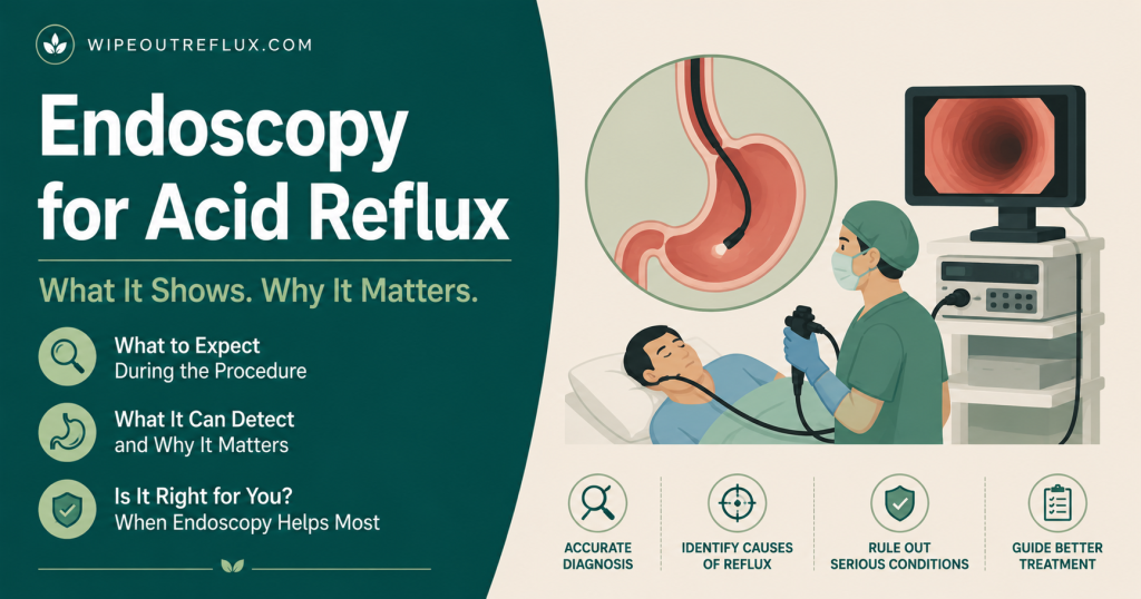
An upper endoscopy for reflux takes about five to ten minutes. You’ll be sedated for it in most US practices, you won’t remember it, and the part people dread — the tube going down — is the part you’re least likely to be aware of. Start to finish, including check-in and recovery, budget two to three hours at the facility, and arrange a ride home.
The procedure itself is genuinely straightforward. What catches people off guard is the result: for most people with reflux symptoms, the endoscopy comes back normal. That is not a mistake, it doesn’t mean your symptoms are imagined, and it doesn’t mean you don’t have reflux. It’s the expected outcome, and understanding why beforehand will save you a lot of anxiety afterwards.
Below is the whole thing step by step — the referral, the prep, the sedation choice, what the gastroenterologist is looking at, why they take biopsies even when everything looks fine, and how to read the report you get afterwards. It’s written US-first, with UK and NHS differences flagged where they matter.
Key Takeaways
- The procedure takes 5–10 minutes; the full visit runs two to three hours including prep and recovery.
- You’ll fast from midnight, or at least 6–8 hours for food and 2 hours for clear liquids.
- US practice is near-universal sedation, often propofol. In the UK you’ll usually be offered a choice between throat spray alone and light sedation.
- Most people with reflux symptoms have a completely normal endoscopy — the majority of GERD is “non-erosive,” meaning no visible damage.
- Biopsies are routine even when the lining looks normal, mainly to check for Barrett’s oesophagus, eosinophilic oesophagitis and H. pylori.
- Guidelines don’t recommend endoscopy for typical reflux symptoms without alarm features — an 8-week PPI trial comes first.
- Alarm features that do warrant prompt endoscopy: trouble swallowing, painful swallowing, unintended weight loss, vomiting, anaemia or signs of bleeding.
- Endoscopy is a poor test for silent reflux (LPR) specifically — a normal scope rules out very little in throat-based reflux.
- Complications are uncommon — reported rates range from about 1 in 200 to 1 in 10,000 depending on how they’re counted — and sedation accounts for a large share of them.
Step 1: The Referral — and Whether You Need One at All
Before the practical steps, it’s worth knowing whether you should be having this test, because a lot of people are scoped who don’t strictly need to be, and a lot of people who should be scoped put it off.
Current US guidance is fairly clear. For someone with classic heartburn and regurgitation, no alarm features, and no particular risk factors, the recommended first step is an eight-week trial of a once-daily PPI taken before a meal — not an endoscopy Katz et al., The American Journal of Gastroenterology, 2022. Endoscopy enters the picture when that trial fails, when alarm features are present, or when there’s reason to screen for Barrett’s oesophagus.
The alarm features worth knowing, because they change the urgency of everything:
- Dysphagia — food sticking, or a sense that it isn’t going down properly.
- Odynophagia — pain on swallowing.
- Unintentional weight loss — weight coming off without you trying.
- Gastrointestinal bleeding — vomiting blood, or black tarry stools.
- Anaemia — particularly iron-deficiency anaemia found on a blood test.
- Persistent vomiting.
If you have any of those alongside reflux, endoscopy should happen promptly rather than after a medication trial. That isn’t a reason to panic — most of these turn out to be benign — but it is a reason not to wait.
There’s also a screening rationale. The 2022 Barrett’s guideline suggests a single screening endoscopy for people with chronic reflux symptoms plus three or more additional risk factors, the list being male sex, age over 50, White race, smoking, obesity, and a first-degree relative with Barrett’s or oesophageal adenocarcinoma Shaheen et al., The American Journal of Gastroenterology, 2022. If that describes you and nobody has mentioned it, it’s a fair thing to raise.
UK note. On the NHS you’ll typically be referred by your GP, and the wait for a routine gastroscopy varies considerably by trust. If alarm features are present you should go onto an urgent suspected-cancer pathway with a much shorter target. It’s worth asking explicitly which pathway you’ve been put on, because it tells you how the referrer has categorised your symptoms.
Step 2: Preparing — Fasting, Medications and Logistics
Fasting. The stomach needs to be empty so the gastroenterologist can see the lining, and so nothing can come back up while you’re sedated. Standard instruction is nothing to eat for at least six to eight hours beforehand — usually “nothing after midnight” for a morning appointment — and clear liquids stopped at least two hours before. Follow the specific times your unit gives you rather than these general ones; they vary, and turning up having eaten usually means cancellation.
PPIs. This one matters more than most people realise and is easy to get wrong. If the purpose of the scope is diagnostic — establishing whether you actually have reflux disease, or screening for Barrett’s — guidelines advise doing it off PPI therapy, typically after two to four weeks without, because acid suppression heals the very changes the test is looking for. Scoping someone on a PPI can turn erosive oesophagitis invisible. But do not stop a PPI on your own initiative before a scope; ask the referring clinician what they want, because stopping abruptly brings its own problems, which I’ve covered in getting off PPIs and acid rebound.
Other medications. Blood thinners, diabetes medication and insulin usually need specific instructions. Most other regular medication can be taken with a small sip of water. Ask ahead rather than assuming.
Logistics. If you’re having sedation you cannot drive afterwards, and most units won’t discharge you into a taxi alone — you need a responsible adult to accompany you home. Clear the rest of the day. Sedation leaves plenty of people foggy for longer than they expect.
Step 3: Arriving and Getting Ready
You’ll check in, change into a gown, and go through the consent conversation with a nurse and usually the endoscopist. They’ll confirm your fasting status, allergies, medications and medical history, and ask about loose teeth or dental work, since the mouthguard sits against your teeth.
A cannula goes into the back of your hand or arm for sedation. You’ll have a blood pressure cuff, a finger probe monitoring your oxygen, and often nasal oxygen. Someone will spray a local anaesthetic into the back of your throat, which tastes bitter and makes your throat feel oddly thick and numb — that sensation is normal and wears off within an hour or so.
This preparation stage takes longer than the procedure itself. That’s normal and not a sign anything is wrong.
Step 4: The Sedation Decision
This is the part people have the most anxiety about, and there’s a genuine choice to understand.
In the US, sedation is close to universal. Most units use either moderate (“conscious”) sedation with a benzodiazepine such as midazolam plus an opioid like fentanyl, or deep sedation with propofol, increasingly administered by an anaesthesia professional. With propofol you’re fully asleep and will have no memory whatsoever. With moderate sedation you’re drowsy and relaxed, may be vaguely aware, and typically remember little or nothing due to the amnesic effect of midazolam.
In the UK, you’ll usually be offered a real choice: throat spray alone, or throat spray plus light intravenous sedation. Throat spray alone means you’re awake throughout, which sounds worse than most people find it — the advantage is no cannula, no recovery period, no escort needed and you can drive yourself home and return to work. Sedation means a more comfortable experience but the whole day is written off. Neither is wrong. If you’re anxious, sedation is a reasonable choice and you should say so rather than feeling you ought to tough it out.
Whichever route, the sedation used for a diagnostic gastroscopy is light by surgical standards. It isn’t general anaesthesia, and you keep breathing on your own throughout.
Step 5: The Procedure Itself
You’ll lie on your left side with your knees slightly bent. A plastic mouthguard goes between your teeth to protect them and hold the scope steady. If you’re having sedation, it goes in now, and for most people that’s the last thing they remember.
The endoscope is a flexible tube about 9 to 10mm across — roughly the width of your little finger, and thinner than most people picture — carrying a light, a camera and a channel for instruments. The endoscopist passes it over your tongue and asks you to swallow once as it reaches the back of the throat — that swallow opens the upper oesophageal sphincter and lets the scope through. This is the only genuinely uncomfortable moment, and it lasts a second or two.
From there the scope passes down the oesophagus into the stomach and on into the first part of the small intestine, the duodenum — which is why the formal name is oesophagogastroduodenoscopy, or EGD. Air or carbon dioxide is gently insufflated to open the folds so the lining can be inspected properly. That’s what causes the bloating and burping afterwards; carbon dioxide is absorbed faster and leaves you less uncomfortable, so it’s worth asking whether your unit uses it.
You can still breathe normally the entire time. The scope is in your oesophagus, which is behind your windpipe, not in it. Everyone worries about this and it’s worth saying plainly.
Expect to gag or retch a little if you’re not deeply sedated. It’s a reflex, not a sign of a problem, and the team sees it constantly.
Step 6: What the Endoscopist Is Actually Looking At
While the scope is in, several specific things are being assessed:
- The oesophageal lining — looking for breaks in the mucosa, which is what erosive oesophagitis is. If present, it’s graded by the Los Angeles classification. Grade A is a break of 5mm or less that doesn’t extend between the tops of two mucosal folds; grade D is a break involving three-quarters or more of the circumference. Worth knowing how those grades are read: grade C or D is considered conclusive evidence of reflux disease, grade B can be diagnostic alongside typical symptoms and a PPI response, but grade A on its own is not enough to confirm GERD — minor breaks turn up in people without it.
- The gastro-oesophageal junction — where the oesophagus meets the stomach, and whether the lining has changed colour and texture in the way that suggests Barrett’s.
- A hiatal hernia — whether the top of the stomach has slid up through the diaphragm, which is common in reflux and affects how it’s managed. More in hiatal hernia and reflux.
- Rings, furrows and narrowing — the concentric rings and longitudinal furrows that suggest eosinophilic oesophagitis rather than reflux, and any stricture that might explain difficulty swallowing.
- The stomach and duodenum — ulcers, inflammation, and signs of H. pylori infection.
Step 7: Biopsies — Why They’re Taken Even When It Looks Normal
People are often unsettled to hear biopsies were taken, assuming it implies something suspicious was seen. Usually it implies the opposite: several important conditions are invisible to the naked eye and can only be found by looking at tissue under a microscope. Taking the biopsy is painless — the oesophageal lining has no fine touch sensation of that kind — and you won’t feel it even if you’re awake.
Here’s what they’re generally looking for.
Barrett’s oesophagus. Chronic acid exposure can cause the lining of the lower oesophagus to change into a more intestine-like tissue. It’s a protective adaptation, but it carries a small increased risk of oesophageal cancer, which is why it’s worth identifying and monitoring. The endoscopist can often see suspicious-looking tissue, but the diagnosis requires biopsy confirmation of intestinal metaplasia. If you have reflux and Barrett’s is a concern, this is the single most valuable thing the test does. There’s more in can silent reflux cause Barrett’s oesophagus.
Eosinophilic oesophagitis. This is the big one for people whose “reflux” never responds to treatment. EoE is an immune-mediated condition where allergic cells infiltrate the oesophagus, and it produces symptoms that mimic reflux almost exactly — but it doesn’t respond to acid suppression in the same way and needs entirely different treatment. Diagnosis requires at least 15 eosinophils per high-power field on biopsy, alongside symptoms of oesophageal dysfunction. Because the infiltration is patchy, guidelines call for at least six biopsies taken from two or more levels of the oesophagus Dellon et al., The American Journal of Gastroenterology, 2025. Crucially, the oesophagus can look completely normal and still be full of eosinophils, which is exactly why biopsies get taken from a normal-looking scope. If your reflux has never responded properly to a PPI, this is the diagnosis you want ruled out — see eosinophilic esophagitis vs acid reflux.
Helicobacter pylori. Biopsies from the stomach lining check for this bacterium, which causes ulcers and gastritis and can produce symptoms overlapping with reflux. It’s treatable with antibiotics, so finding it changes management immediately.
Coeliac disease. Duodenal biopsies are often taken as well, since coeliac disease can present with vague upper digestive symptoms and is easy to miss otherwise.
Results from biopsies typically take one to three weeks, which is longer than the verbal summary you get on the day. That gap is normal.
Step 8: Recovery and Going Home
If you were sedated, you’ll wake in a recovery area over roughly 30 to 60 minutes while your observations are monitored. If you had throat spray only, you’ll be able to leave much sooner, though you can’t eat or drink until the numbness wears off — usually about an hour — because a numb throat makes choking easier.
What to expect for the rest of the day:
- A sore or scratchy throat for a day or so. Normal, and usually mild.
- Bloating and burping from the air used during the procedure. This settles within a few hours.
- Grogginess if you were sedated. No driving, no alcohol, no signing anything important, and ideally no important conversations for 24 hours.
- Patchy memory of the conversation immediately afterwards — which is why you’re given written results and why having someone with you helps.
Eat lightly at first. Something soft and bland is more comfortable than a full meal, and it’s worth avoiding your usual reflux triggers for the rest of the day while your throat settles.
When to call for help. Serious complications are uncommon, but you should contact the unit or seek urgent care for severe or worsening chest or abdominal pain, difficulty breathing, fever, vomiting blood, or black tarry stools. Overall, adverse events from diagnostic upper endoscopy are uncommon. Large series report rates ranging from around 1 in 200 to 1 in 10,000, the spread reflecting how broadly “adverse event” is defined and how sick the patients were. Perforation is rarer still, somewhere between 1 in 2,500 and 1 in 11,000. Notably, cardiopulmonary events related to sedation account for as much as 60% of the total — which is to say a good deal of the risk comes from being sedated rather than from the scope Ben-Menachem et al., Gastrointestinal Endoscopy, 2012.
Step 9: Understanding a Normal Result
Here’s the part that causes the most confusion, and it deserves a proper explanation rather than a line in a discharge letter.
Most people with genuine reflux symptoms have a normal-looking endoscopy. Depending on the population studied, somewhere between half and three-quarters of people with typical reflux symptoms show no visible erosions at all. This has a name — non-erosive reflux disease, or NERD — and it’s the most common form of GERD, not an exception to it.
Why? Because endoscopy detects damage, not reflux. Whether reflux causes visible erosions depends on how acidic the refluxate is, how long it sits there, how well your oesophagus clears it, and how resistant your mucosa happens to be. Plenty of people reflux frequently and symptomatically without ever developing breaks in the lining. Their oesophagus may also simply be more sensitive — registering as painful an amount of acid that someone else wouldn’t notice.
So a normal endoscopy tells you something genuinely useful: you don’t have erosive oesophagitis, a stricture, Barrett’s, EoE, an ulcer or a tumour. That’s a lot of reassurance. What it does not tell you is whether you have reflux. If symptoms persist despite a normal scope, the next step is usually pH or pH-impedance testing, which measures reflux directly rather than inferring it from damage — covered in the Bravo pH test and how acid reflux is diagnosed. Oesophageal manometry may follow if a motility problem is suspected.
Endoscopy and Silent Reflux: A Specific Warning
If your symptoms are throat-based — chronic throat clearing, a lump sensation, hoarseness, post-nasal drip, a cough that won’t quit — you need to know that upper endoscopy is a weak test for what you have.
Silent reflux, or LPR, involves small amounts of refluxate, often gaseous and often containing pepsin, reaching the throat and larynx. The oesophagus may be entirely undamaged because the material passes through quickly, while the far more delicate laryngeal tissue is irritated by comparatively tiny exposures. So a normal gastroscopy is exactly what you’d expect in LPR, and it rules out very little.
The laryngoscopy that ENT surgeons perform isn’t much more definitive on its own. The laryngeal signs attributed to reflux — redness, swelling, ventricular obliteration, pseudosulcus — turn out to be common in healthy people too, and interpretation varies considerably between clinicians, which gives these signs poor specificity Lechien et al., Otolaryngology–Head and Neck Surgery, 2023. That doesn’t mean the test is worthless — it’s important for excluding other causes of hoarseness, including ones that need ruling out promptly — but a diagnosis of LPR shouldn’t rest on the scope picture alone.
For LPR the more informative tests tend to be symptom scoring with the Reflux Symptom Index, pharyngeal pH monitoring such as Restech, or salivary pepsin testing like Peptest. If you go in for a gastroscopy expecting it to confirm silent reflux, you’re likely to come out frustrated. Going in knowing it’s there to exclude other things is a much healthier expectation.
Conclusion
An endoscopy is a short, safe, well-tolerated procedure, and the anticipation is reliably worse than the event. Fast as instructed, sort out your ride home, ask what your unit’s sedation options are, and be clear with the endoscopist beforehand about whether you should be off your PPI. Then expect the most likely outcome: a normal-looking oesophagus, some biopsies pending, and a letter confirming there’s nothing structurally wrong.
That normal result is worth more than it feels like at the time. It rules out the things that genuinely need ruling out, and it reframes the problem — because if there’s no damage to heal, the question stops being “how do I heal this” and becomes “how do I stop the reflux happening in the first place.” That’s a dietary and mechanical question far more than a pharmaceutical one, and it’s where the real progress usually gets made.
That’s exactly the ground the Wipeout Diet Plan covers — a structured way to reduce the reflux events themselves rather than just suppressing the acid in them. I built it primarily around LPR and silent reflux, the throat-based form where scopes come back clear most often and medication disappoints most reliably, but because it works on the same underlying mechanisms it does the job just as well for GERD and everyday heartburn. And if you want somewhere concrete to start while you’re waiting on biopsy results, the Wipeout Food Reference Guide is the essential companion — the full list of which foods and drinks are safe for acid reflux and LPR, with their actual pH values, so you’re working from numbers rather than guesswork.
Frequently Asked Questions
Does an endoscopy hurt?
No. With sedation you won’t be aware of it at all. With throat spray only, the swallow as the scope passes the back of the throat is briefly uncomfortable and you may gag, but it isn’t painful — the oesophagus doesn’t register that kind of sensation. Most people afterwards say the anticipation was far worse than the procedure.
How long does it take?
The scope itself is usually five to ten minutes. Add biopsies and it might reach fifteen. Including check-in, preparation and recovery, plan on two to three hours at the facility.
Will I be asleep?
In the US, usually yes — either deeply sedated with propofol, or moderately sedated with midazolam and fentanyl, which typically wipes your memory of it. In the UK you’ll usually be offered a choice between throat spray alone, where you’re fully awake, and light sedation.
Can I drive myself home?
Only if you had throat spray and no sedation. With any intravenous sedation you cannot drive for 24 hours and will need a responsible adult to take you home.
Should I stop my PPI before the endoscopy?
Ask the clinician who referred you — don’t decide alone. If the scope is diagnostic or screening for Barrett’s, they’ll often want you off it for two to four weeks, because acid suppression heals the damage the test is looking for and can mask erosive oesophagitis. But stopping abruptly can cause rebound symptoms, so it needs to be planned.
What if my endoscopy is normal but I still have symptoms?
That’s the most common outcome, not a failure of the test. Most reflux is non-erosive, meaning symptoms without visible damage. It rules out erosions, Barrett’s, EoE, strictures and ulcers. If symptoms continue, pH or pH-impedance monitoring measures reflux directly and is the usual next step.
Why did they take biopsies if everything looked fine?
Because several conditions are invisible to the naked eye. Eosinophilic oesophagitis in particular can occur in an oesophagus that looks completely normal, and Barrett’s needs tissue confirmation. Biopsies from a normal-looking scope are routine good practice, not a sign something worrying was spotted.
How long do results take?
You’ll usually get a verbal summary and a written report of the visual findings the same day, though sedation means many people don’t retain the conversation. Biopsy results take longer — typically one to three weeks.
Can an endoscopy diagnose silent reflux?
Not reliably. LPR usually leaves the oesophagus undamaged, so a normal scope is expected and rules out very little. Endoscopy is valuable for excluding other conditions, but LPR is better assessed with symptom scoring, pharyngeal pH monitoring or salivary pepsin testing.
Is endoscopy dangerous?
Complications are uncommon. Reported rates span roughly 1 in 200 to 1 in 10,000, depending on how adverse events are defined and how unwell the patients were, with perforation somewhere between 1 in 2,500 and 1 in 11,000. Cardiopulmonary events linked to sedation make up as much as 60% of the total, so much of the risk attaches to being sedated rather than to the scope itself — worth weighing if you’re offered throat spray alone. For most people the risk is low relative to the information gained.
Research & References
- Katz et al., The American Journal of Gastroenterology, 2022 — ACG clinical guideline for the diagnosis and management of gastro-oesophageal reflux disease, recommending an 8-week empiric once-daily PPI trial for patients with typical symptoms and no alarm features, prompt endoscopy where alarm features are present, and diagnostic endoscopy performed off PPI therapy.
- Shaheen et al., The American Journal of Gastroenterology, 2022 — Updated ACG guideline on Barrett’s oesophagus, suggesting a single screening endoscopy for patients with chronic reflux symptoms plus three or more additional risk factors (male sex, age over 50, White race, smoking, obesity, or a first-degree relative with Barrett’s or oesophageal adenocarcinoma).
- Dellon et al., The American Journal of Gastroenterology, 2025 — ACG clinical guideline on eosinophilic oesophagitis, setting the histological diagnostic threshold at 15 eosinophils per high-power field alongside symptoms of oesophageal dysfunction, and recommending at least six biopsies from two or more oesophageal levels because the infiltrate is patchy.
- Ben-Menachem et al., Gastrointestinal Endoscopy, 2012 — ASGE Standards of Practice Committee guideline on adverse events of upper GI endoscopy. Large series report overall adverse event rates of 1 in 200 to 1 in 10,000 and mortality from none to 1 in 2,000; cardiopulmonary events 1 in 170 to 1 in 10,000; perforation 1 in 2,500 to 1 in 11,000. Cardiopulmonary events related to sedation and analgesia account for as much as 60% of upper GI endoscopy adverse events.
- Lechien et al., Otolaryngology–Head and Neck Surgery, 2023 — Study of the sensitivity, specificity and predictive values of laryngopharyngeal reflux symptoms and signs in clinical practice, concluding that LPR symptoms and signs show low specificity and negative predictive value; the combined non-endoscopic sign cluster reached only 47.2% specificity, though several individual signs performed better in isolation.

David Gray
Content Researcher & Author
David Gray founded Wipeout Reflux to address a critical gap in reflux management. His research synthesizes over 100 peer-reviewed studies on laryngopharyngeal reflux (LPR), pepsin biology, and GERD pathophysiology. For LPR specifically—a condition most physicians misdiagnose—his work focuses on pepsin reactivation and why standard PPI therapy fails most patients. He develops evidence-based protocols targeting root causes of both LPR and GERD, integrating emerging research on sphincter dysfunction, dietary interventions, and newer clinical approaches. Wipeout Reflux represents practical application of clinical science for patients seeking real solutions.