
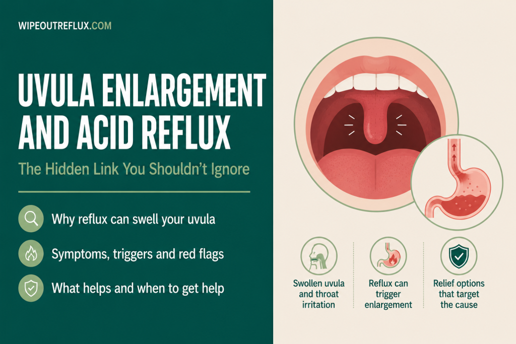
Most people notice the uvula — that small, teardrop-shaped piece of tissue hanging at the back of the throat — only when it’s causing a problem. If yours looks larger, redder, or more swollen than usual and you have acid reflux or LPR, the connection isn’t a coincidence.
Uvula swelling and redness (uvulitis) is a documented and, in LPR, surprisingly specific sign of reflux reaching the upper throat. A major clinical study of 403 LPR patients found that uvula erythema and oedema carries a specificity of 97.1% for laryngopharyngeal reflux — meaning when an ENT clinician sees these changes in the uvula, LPR is almost certainly the cause. That makes it one of the most diagnostically reliable physical signs of the condition.
The reason is anatomical and straightforward. The uvula hangs directly in the path of nighttime reflux. During sleep, when the throat is horizontal and defences are down, acid and pepsin rising from the stomach encounter the uvula first. Over time, repeated exposure inflames, swells, and can significantly elongate the tissue. The good news — and this is well documented clinically — is that uvula enlargement from reflux is reversible. Treat the reflux effectively, and the uvula typically returns to normal size within weeks.
Key Takeaways
- Uvula erythema and oedema has a specificity of 97.1% for LPR in clinical studies — one of the highest specificities of any visible LPR sign, meaning it is a particularly reliable indicator when present.
- The uvula is especially vulnerable to reflux damage because of its position: it hangs directly into the path of gastric contents that reach the throat during sleep, when swallowing and protective reflexes are suppressed.
- Pepsin — not just acid — is the primary driver of tissue damage. Pepsin is taken up by mucosal cells and continues to cause inflammatory injury at the pH levels typically present in the upper throat.
- LPR-related uvula enlargement is reversible with appropriate anti-reflux treatment. Clinicians experienced in LPR report that uvula size can return to normal within weeks of reflux being controlled.
- A swollen uvula from reflux can contribute to snoring and obstruction — and conversely, snoring and obstructive sleep apnoea worsen LPR through negative airway pressure that draws stomach contents upward, creating a self-reinforcing cycle.
- Other causes of uvula swelling — infection, allergy, angioedema, and dehydration — need to be considered alongside reflux, particularly when swelling is sudden, severe, or accompanied by breathing difficulty.
- A rapidly enlarging uvula with difficulty breathing or swallowing is an emergency. Reflux-related uvula changes are gradual; sudden severe swelling of the uvula always requires urgent medical attention.
What the Uvula Is and Why It’s Particularly Vulnerable to Reflux
The uvula (palatine uvula) is a small, finger-like projection of soft tissue that hangs from the midpoint of the soft palate at the back of the mouth. It’s composed of connective tissue, muscle fibres, and mucous glands, and it serves several functions — contributing to speech, helping seal the nasal passages during swallowing, triggering the gag reflex when touched, and producing saliva and mucus that help lubricate the oropharynx.
That last function — secreting lubricating mucus — is part of what makes the uvula an active participant in oropharyngeal defence. But it also makes the uvula’s tissue inherently rich in glandular mucosa, and glandular mucosal tissue is significantly more reactive to chemical irritants than the tougher, stratified squamous epithelium lining most of the throat. When pepsin and acid reach the uvula, the response is faster and more pronounced than in tissue adapted to withstand more abrasion.
The uvula’s position is the other key vulnerability. It hangs at the precise back of the oral cavity, directly above the oropharynx — the part of the throat that becomes the first landing zone for reflux material that passes the upper oesophageal sphincter. During the day, swallowing every 30–60 seconds continuously clears material from this region. During sleep, swallowing drops to nearly nothing, the supine position eliminates gravitational protection, and the soft palate and uvula are directly exposed to anything that reaches the upper throat. For LPR sufferers, this means the uvula is bathed in pepsin-containing refluxate for hours at a time, night after night.
How LPR Causes Uvula Swelling and Redness: The Mechanism
The mechanism of reflux-driven uvula damage follows the same pathway as LPR damage elsewhere in the throat — but the uvula, for the anatomical reasons above, may receive more intense and sustained exposure than the vocal cords or posterior commissure in some patients.
When gastric contents reach the oropharynx during a reflux event, the primary chemical aggressors are pepsin and acid working together. Pepsin is the dominant damaging agent in the upper throat. At the acidic pH typically found in refluxate (pH 2–4), pepsin is fully active and capable of directly digesting mucosal surface proteins. But — and this is critical for understanding why LPR damage is so persistent — pepsin is also taken up by epithelial cells and remains stable within them at a wide pH range. Even as the ambient pH in the throat rises back toward neutral after a reflux event, intracellular pepsin can be reactivated by the next exposure to acidity, whether from food, drink, or another reflux event.
This means that every reflux event adds to a cumulative pepsin load in the uvular tissue. Research using animal models has confirmed that repeated exposure to pepsin and even weakly acidic reflux causes progressive breakdown of E-cadherin — the protein that maintains the integrity of the epithelial barrier — and widening of the spaces between mucosal cells. Once the barrier is disrupted, inflammatory mediators penetrate more easily, the tissue becomes hyperaemic (engorged with blood) and oedematous (filled with fluid), and the uvula swells, elongates, and takes on a reddened appearance.
This is the pathological picture that ENT clinicians describe when they refer to uvula erythema and oedema as an LPR sign. It’s not infection, it’s not allergy — it’s the local inflammatory response to chronic pepsin-mediated tissue injury.
The full LPR mechanism — including how pepsin reaches and damages the upper throat more broadly — is covered in the complete guide to LPR. The uvula is one specific site within a wider pattern of upper aerodigestive tract involvement.
Uvula Changes as a Diagnostic Sign: What the Numbers Mean
What makes uvula erythema and oedema clinically important — beyond being something a patient notices in the mirror — is its diagnostic utility, which is considerably higher than most LPR signs.
A prospective clinical study of 403 LPR patients (confirmed by hypopharyngeal-oesophageal impedance pH monitoring) and 144 healthy controls found that uvula erythema and oedema had a specificity of 97.1–97.2% for LPR. To put that in context: specificity measures how reliably a sign is absent in people who don’t have the disease. A 97% specificity means that when an ENT sees this finding, it is almost exclusively present in LPR patients, not in healthy individuals.
By comparison, many of the most common LPR signs — throat clearing, hoarseness, posterior commissure hypertrophy — have much lower specificity, because they can appear in healthy individuals or in response to other causes. The uvula finding stands out as one of only three clinical signs (alongside epiglottis erythema and interarytenoid granulatory tissue) that reached this very high specificity threshold.
The study also found that combining uvula erythema/oedema with throat clearing, heartburn, globus sensation, and anterior pillar erythema produced the highest diagnostic combination overall — achieving 98.8% sensitivity and 33.3% specificity, with a positive predictive value of 94.3%.
This is why experienced LPR clinicians often note the uvula specifically during examination. If you’ve had a throat examination where an ENT mentioned your uvula looked swollen or red, and you have LPR symptoms, that observation should be taken seriously as a clinically meaningful finding rather than an incidental comment.
Understanding the full range of LPR signs — many of which occur without any heartburn — is important context here. The LPR symptoms guide covers the full clinical picture, including signs that are commonly missed.
The Snoring and Sleep Apnoea Connection
The relationship between an enlarged uvula, snoring, and obstructive sleep apnoea (OSA) intersects with reflux in a way that creates a self-reinforcing cycle — and understanding it helps explain why some patients find their uvula progressively more enlarged despite treating reflux, or why reflux seems to worsen alongside snoring even when diet is managed carefully.
An enlarged uvula from LPR-related swelling can itself contribute to upper airway obstruction. A longer, oedematous uvula creates more soft tissue in the oropharynx, increasing resistance to airflow during sleep. This can trigger snoring — the sound of that tissue vibrating in the partially narrowed airway — and in more significant cases, contribute to obstructive episodes where the airway collapses partially or completely.
Snoring and OSA then feed back into reflux through a different mechanism. The negative intrathoracic pressure generated during obstructed breathing events during sleep — the effort of breathing against a partially blocked airway — physically draws stomach contents upward toward the oesophagus and throat. This is the same “suction” mechanism that makes OSA such a consistent risk factor and worsening factor for both GERD and LPR. Genetic analysis using Mendelian randomisation has confirmed that the relationship is genuinely bidirectional and causal: GERD elevates the risk of OSA and snoring, and OSA independently worsens GERD through these airway pressure dynamics.
The practical implication is that a patient with LPR who snores may have a particularly vicious cycle operating: LPR inflames the uvula → the enlarged uvula worsens snoring → snoring worsens LPR → which further inflames the uvula. Breaking this cycle requires addressing both the reflux and the sleep-disordered breathing rather than treating each in isolation. The article on sleep apnoea and acid reflux covers this bidirectional relationship in depth.
Importantly, uvula surgery (uvulopalatopharyngoplasty, or UPPP — often performed to reduce snoring or treat sleep apnoea) should not be the first intervention when LPR is the underlying cause of uvula enlargement. Removing or reducing the uvula surgically while the reflux continues will not address the root cause, and the surrounding tissue will remain inflamed and potentially obstructed. Anti-reflux treatment first, allowing the uvula to return to normal size, is the appropriate sequence — with surgical assessment only if relevant obstruction persists after reflux is adequately controlled.
Other Causes of Uvula Swelling: What to Consider
Reflux is a well-documented cause of uvula swelling — but it’s not the only one, and some other causes require different and more urgent management. It’s important to have the full picture, particularly in distinguishing chronic reflux-driven changes from acute or potentially dangerous swelling.
Infection (uvulitis): Bacterial and viral infections — strep throat, mononucleosis, other upper respiratory infections — are among the most common causes of acute uvula swelling in the general population. Infection-related swelling typically comes with fever, sore throat, tonsillar exudate, and generalised throat inflammation rather than the isolated posterior throat redness pattern of LPR.
Allergic reaction: Allergies to foods, medications, or inhaled allergens can cause uvula swelling as part of a broader allergic oropharyngeal response. Seasonal allergies, dust mite sensitivity, and allergic rhinitis all cause throat inflammation that may include the uvula.
Angioedema: This is the most urgent differential diagnosis for uvula swelling. Angioedema — swelling of the deeper layers of the throat tissue driven by histamine release or other mechanisms — can cause rapidly progressive uvula swelling that threatens the airway. Hereditary angioedema (HAE) is a rare genetic condition that produces recurrent unexplained episodes of throat, face, and limb swelling without obvious trigger. ACE inhibitor medications (commonly prescribed for blood pressure) can cause angioedema as a side effect, and this is a genuine emergency when it affects the throat.
Dehydration and alcohol: Significant dehydration or alcohol consumption can cause transient uvula swelling, particularly if the uvula dries out overnight — this tends to resolve with rehydration.
Mechanical irritation: Intubation, endoscopy, tonsillectomy, and dental procedures can all cause post-procedural uvula swelling that typically resolves over days.
The key distinguishing feature of reflux-related uvula changes is that they are gradual, chronic, and associated with other LPR symptoms — throat clearing, hoarseness, globus sensation, post-nasal drip — rather than sudden, painful, or accompanied by fever or breathing difficulty. If uvula swelling is new, rapidly worsening, or accompanied by any of the features in the next section, it needs prompt medical assessment.
When Uvula Swelling Needs Urgent Medical Attention
Most reflux-related uvula changes develop slowly over time and don’t constitute an emergency. But uvula swelling from any cause can, in significant cases, progress to airway compromise — and the following presentations require immediate medical evaluation rather than a wait-and-see approach:
- Difficulty breathing or stridor (a high-pitched breathing sound) — any breathing difficulty alongside throat swelling is an emergency
- Rapid or sudden onset of uvula swelling, particularly without obvious cause — this pattern suggests angioedema rather than reflux, and can escalate quickly
- Severe difficulty swallowing, particularly with drooling, as this suggests significant oropharyngeal obstruction
- High fever with uvula swelling — suggests epiglottitis, peritonsillar abscess, or another serious infection that needs urgent ENT evaluation
- Uvula swelling alongside facial, lip, or tongue swelling — strongly suggests angioedema; if you take an ACE inhibitor, this is a known serious reaction, call emergency services immediately
- Progressive worsening despite no infection and no reflux flare — any unexplained progressive throat swelling should be assessed by an ENT or emergency physician
Reflux does not produce the sudden-onset, rapidly-progressive swelling associated with angioedema or severe infection. If your uvula swelling fits the slow, chronic pattern alongside known LPR symptoms, reflux management is the appropriate next step. If there is any doubt about acuity or cause, err toward medical review.
Can a Swollen Uvula From Reflux Return to Normal?
Yes — and this is one of the more encouraging aspects of this particular presentation. Clinicians with significant LPR experience, including Dr Jamie Koufman whose work on respiratory reflux and LPR is widely cited, have documented that uvula enlargement driven by reflux is reversible when reflux is adequately controlled. In some cases the uvula returns to normal size within a matter of weeks on a strict anti-reflux programme.
The tissue changes involved — oedema (fluid accumulation) and inflammatory hyperaemia — are not the same as permanent structural changes like fibrosis or scarring. As long as the chronic pepsin exposure driving the inflammation is removed, the mucosal tissue can recover. This is consistent with what happens in other LPR-affected sites: posterior commissure hypertrophy, laryngeal oedema, and supraglottic inflammation all improve with effective reflux management, though timelines vary depending on severity and duration of exposure.
Cases where the uvula is severely elongated — particularly those associated with long-standing OSA and snoring alongside reflux — may take considerably longer, with months of controlled reflux required before the structural changes in the soft palate and uvula fully resolve. In those cases, surgical assessment for the residual structural component may eventually be appropriate, but only once reflux has been optimised and the inflammatory contribution is eliminated.
The implication for management is important: if an ENT proposes uvula surgery for snoring or obstruction in a patient with known or suspected LPR, a serious and sufficiently long trial of reflux control should come first. Removing inflamed, reflux-swollen tissue while the underlying cause continues is unlikely to produce lasting improvement.
What to Do If You Have LPR and Uvula Swelling
The management follows directly from the mechanism: the priority is reducing the frequency and reach of reflux events so that pepsin stops reaching the uvula night after night.
Dietary changes are the foundation. The foods and drinks that most reliably worsen LPR — acidic, spicy, fatty, and caffeinated — should be reduced or eliminated during the active treatment phase. This isn’t permanent restriction, but the healing phase requires keeping the pepsin burden as low as possible. The LPR foods to avoid guide covers the most impactful changes with the mechanism behind each one.
Address nighttime reflux specifically. Because the uvula is particularly exposed during sleep, nighttime management is disproportionately important. Eating at least three hours before bed, sleeping on your left side, and elevating the head of the bed by 15–20 cm all reduce the frequency of nocturnal reflux events reaching the throat. The guide to sleeping position for silent reflux covers the specifics.
Use alginates, particularly at bedtime. Gaviscon Advance taken before lying down forms a raft that covers the gastro-oesophageal junction throughout the night, reducing the material available to reach the throat. The alginate also directly inhibits pepsin — reducing the proteolytic activity of any refluxate that does reach the uvula. This is a particularly important treatment step when the throat and upper structures are the primary site of damage.
Investigate and address snoring or sleep apnoea. If you snore regularly or have any features of OSA (waking unrefreshed, episodes where breathing stops, daytime sleepiness despite adequate sleep), assessment and management of the airway component is important. Untreated OSA worsens LPR through negative pressure mechanics and will work against your reflux management efforts regardless of how carefully you manage diet.
Work with an ENT who understands LPR. Uvula findings in LPR are well documented in the research but are not yet universally recognised in all clinical settings. An ENT with specific expertise in LPR — who will look for the full pattern of throat findings including uvula changes — is more likely to put the picture together correctly and to sequence treatment appropriately before recommending any surgical intervention.
Frequently Asked Questions
Can acid reflux cause a swollen uvula?
Yes. LPR (laryngopharyngeal reflux, or silent reflux) is a documented cause of uvula swelling and redness. Pepsin and acid reaching the throat during reflux — particularly during sleep — cause progressive inflammatory changes in the uvula, producing oedema and erythema. Uvula erythema/oedema has a 97% specificity for LPR in clinical studies, making it one of the most reliable visible signs of the condition.
Why does my uvula look bigger when I wake up?
Morning uvula swelling in the context of reflux is characteristic of nighttime LPR. During sleep, swallowing stops, the horizontal position removes gravitational protection, and pepsin-containing refluxate can reach and bathe the uvula for prolonged periods. Waking up with a swollen, irritated uvula that improves during the day is a common pattern in LPR patients, particularly before the condition is adequately treated. Mouth breathing and snoring during sleep are contributing factors.
What does an enlarged uvula feel like with LPR?
People with LPR-related uvula swelling often describe a feeling of something hanging at the back of the throat, a persistent tickling or gagging sensation, the urge to clear the throat, difficulty swallowing — particularly the sensation that the uvula is touching the tongue or triggering the gag reflex when eating. Some describe it as the classic globus sensation, though this can have multiple overlapping causes in LPR.
Will my uvula go back to normal if I treat my reflux?
Yes, in most cases. Clinical experience in LPR management consistently shows that uvula enlargement driven by reflux resolves when reflux is adequately controlled. The inflammatory oedema is not permanent structural damage, and the tissue can recover once the chronic pepsin exposure is removed. Some cases — particularly those with long-standing severe LPR and associated snoring — may take longer, but resolution is the expected outcome of successful reflux management.
Should I have surgery for an enlarged uvula caused by reflux?
Not before trying anti-reflux treatment first. Surgery on a reflux-inflamed uvula without controlling the underlying cause is unlikely to produce lasting improvement. The standard clinical recommendation is to manage the reflux adequately first — typically for a minimum of several months — and assess the uvula and airway once inflammation has resolved. Surgical assessment then becomes relevant only if structural obstruction persists after the inflammatory component has been addressed.
Can uvula swelling from reflux affect breathing or sleep?
In significant cases, yes. A substantially enlarged uvula adds soft tissue to the oropharynx, increasing resistance to airflow during sleep and contributing to snoring or worsening existing obstructive sleep apnoea. This is one reason why LPR-related uvula enlargement, snoring, and OSA tend to occur together, and why treating the reflux can improve all three simultaneously — as the uvula returns to normal size, the airway opens and sleep quality improves.
How is uvula swelling from reflux different from infection?
Reflux-related uvula changes are gradual, chronic, and typically worse in the morning. They are associated with the broader LPR symptom pattern — throat clearing, hoarseness, globus, post-nasal drip — without fever or generalised throat pain. Infectious uvulitis tends to be more acute in onset, accompanied by fever, marked throat pain, and often other signs of infection such as tonsillar swelling or exudate. A sudden onset of uvula swelling with systemic features (fever, difficulty swallowing, breathing changes) always warrants prompt medical review.
Conclusion
Uvula enlargement isn’t a symptom most people associate with acid reflux — but it should be, particularly in anyone with LPR. The uvula’s position directly in the path of nighttime reflux, the susceptibility of its glandular mucosa to pepsin-mediated injury, and the documented 97% specificity of uvula erythema and oedema for LPR all make this a clinically meaningful finding that deserves more attention than it typically gets.
The key message is practical and positive: this is reversible. The swelling isn’t permanent structural change — it’s inflammatory oedema driven by ongoing pepsin exposure. Control the reflux, and the uvula recovers. The dietary foundation for doing that is the same as for LPR management more broadly: reducing the foods and behaviours that drive reflux events, managing nighttime exposure specifically, using alginates at the right times, and addressing any co-existing sleep-disordered breathing that is both worsening LPR and being worsened by it.
If you’re building the dietary side of your LPR management, the LPR diet guide and the Wipeout Diet Plan provide the structured framework — covering not just which foods to avoid but the phased approach to rebuilding a diet that keeps reflux at bay long term. The Wipeout Food Reference Guide is the essential companion — pH values and reflux potential for the foods and drinks you encounter day to day, so every food choice is informed rather than guessed.
Research Sources
[__Lechien JR, Otolaryngology–Head and Neck Surgery, 2023__] — Prospective clinical study of 403 LPR patients (confirmed by hypopharyngeal-oesophageal impedance pH monitoring) and 144 healthy controls; established that uvula erythema/oedema has a specificity of 97.1–97.2% for LPR — one of the three highest-specificity signs in the entire LPR clinical sign repertoire; also identified that combining uvula erythema/oedema with throat clearing, heartburn, globus, and anterior pillar erythema achieves 98.8% sensitivity and 94.3% positive predictive value.
[__Li et al., Frontiers in Medicine, 2025__] — Animal model study directly demonstrating that pepsin and weakly acidic reflux cause progressive laryngopharyngeal mucosal barrier injury; confirmed E-cadherin downregulation correlating with acid strength, widening of intercellular spaces, and penetration of lanthanum staining into epithelial junctions — establishing the molecular mechanism of mucosal damage underlying uvula and laryngeal tissue swelling in LPR.
[__Belafsky et al., Laryngoscope, 2001__] — Original validation study for the Reflux Finding Score (RFS), the standard laryngoscopic assessment tool for LPR; established that posterior laryngeal oedema, erythema, diffuse laryngeal oedema, and other signs including those now recognised to include the uvula can be reliably scored and tracked; demonstrated that RFS changes correlate with treatment response in LPR patients on double-probe pH monitoring.
[__Lechien JR et al., Journal of Clinical Medicine, 2022__] — Comparison study of LPR symptoms and findings in 403 confirmed LPR patients vs clinician-estimated prevalence; confirmed uvula erythema as a sign included in the Reflux Sign Assessment scoring instrument; found that otolaryngologists systematically underestimate the prevalence of certain LPR findings including uvula signs, highlighting the gap between clinical evidence and bedside practice.
[__Lee et al., Laryngoscope Investigative Otolaryngology, 2022__] — Study of 91 participants examining LPR changes after UPPP surgery for OSA; confirmed that OSA severity correlates with both Reflux Symptom Index and Reflux Finding Score; documented that laryngeal LPR findings — including diffuse laryngeal oedema of the type affecting the uvula — are consistently higher in OSA patients; demonstrated that treating OSA can improve LPR scores, supporting the bidirectional relationship between uvula/soft palate inflammation, airway obstruction, and reflux.
[__Tian et al., Frontiers in Neuroscience, 2023__] — Bidirectional two-sample Mendelian randomisation study confirming a causal relationship between GERD and obstructive sleep apnoea, insomnia, and snoring; GERD genetically elevated the risk of OSA (OR 1.07), snoring, and insomnia; conversely, OSA independently elevated the risk of GERD; provided genetic-level evidence for the bidirectional causal relationship relevant to the uvula-snoring-LPR cycle.
[__Lechien JR et al., Frontiers in Medicine, 2021__] — Five-year retrospective case series of 21 patients with atypical LPR presentations confirmed by pH-impedance monitoring; demonstrated that diverse extralaryngeal manifestations including oral, sinonasal, and otological findings resolved after anti-reflux treatment (diet, PPIs, alginates); relevant to the reversibility argument — atypical inflammatory changes driven by LPR are expected to resolve once reflux is controlled.
[__Martinucci et al., World Journal of Gastrointestinal Pharmacology and Therapeutics, 2015__] — Review of LPR diagnosis and treatment; summarised the evidence base for laryngoscopic findings in LPR including posterior laryngeal erythema and oedema; addressed the limitation that many LPR signs have low specificity when examined individually, reinforcing the significance of the uvula’s comparatively high specificity as a clinical sign; reviewed the role of pepsin and acid as primary laryngopharyngeal mucosal aggressors.
David Gray
Content Researcher & Author
David Gray founded Wipeout Reflux to address a critical gap in reflux management. His research synthesizes over 100 peer-reviewed studies on laryngopharyngeal reflux (LPR), pepsin biology, and GERD pathophysiology. For LPR specifically—a condition most physicians misdiagnose—his work focuses on pepsin reactivation and why standard PPI therapy fails most patients. He develops evidence-based protocols targeting root causes of both LPR and GERD, integrating emerging research on sphincter dysfunction, dietary interventions, and newer clinical approaches. Wipeout Reflux represents practical application of clinical science for patients seeking real solutions.